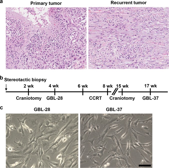
Fig. 1. Isolation and culture of tumor cells from a patient with primary and recurrent glioblastoma.
a Representative images of the H&E-stained sections of the primary and recurrent tumors (original magnification, 50×). b A schematic schedule of the stereotactic biopsy, craniotomy, concurrent chemoradiation therapy (CCRT), and preparation of the patient-derived cells (GBL-28 and GBL-37 cells). wk, week(s) after initial diagnosis and stereotactic biopsy. c Representative images of GBL-28 and GBL-37 cells. Scale bar: 100 μm

