Figure 1.
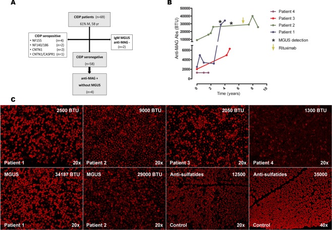
Flowchart of the study population (A). Serial anti-MAG antibody titers during follow-up (B). The asterisks highlight the detection of IgM MGUS in patients 1 and patient 2. The arrow indicates rituximab administration. Immunohistochemistry studies with serum from patients 1–4 showing IgM binding on the myelin sheaths. Immunofluorescence intensity increased in patients 1 and 2 after MGUS detection (C). Staining pattern of patients anti-MAG- sulfatides+ MGUSP used as control are shown. Titers of anti-MAG and anti-sulfatides antibodies are represented. (Anti-IgM, 20x and 40x original magnification). BTU Bühlmann test units; IgM immunoglobulin M; MAG myelin-associated glycoprotein; MGUS monoclonal gammopathy of uncertain significance.

