Figure 5.
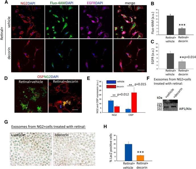
Decorin drives NG2+ cells differentiation into oligodendrocytes through control of RA-associated exosome release via the EGFR–calcium pathway. A, NG2+ cells cultured on a CSPG substrate in the presence of retinal or retinal with decorin for 3 h show a downregulation of EGFR and intracellular Ca2+ (Fluo-4AM) in the decorin-treated cultures. B, C, Quantification of Fluo-4AM and EGFR immunofluorescence in the cultures. D, Expression of OSP in the same cultures. E, Quantification of NG2 and OSP+ cells show that decorin enhances significantly the differentiation of NG2+ cells to oligodendrocytes. Data represent mean of fluorescence intensities in arbitrary units (a.u.) ± SEM calculated from five fields per culture condition from three independent experiments, **p ≤ 0.005, ***p ≤ 0.001, Student's t test. F, Western blot of exosomes isolated from retinal + vehicle- and retinal + decorin-treated cultures with AIP1/Alix antibody. G, β-galactoside staining of RARE LacZ transfected F9 cells showing RA in exosomes isolated from NG2+ cell cultures treated with: vehicle + retinal and decorin + retinal. H, Quantification of RA shown as percentage of LacZ+ cells suggests that decorin prevents the secretion of RA in exosomes. Data are shown as mean ± SEM obtained from five fields per culture treatment from three independent experiments, ***p ≤ 0.001, Student's t test. Scale bars, 50 μm.

