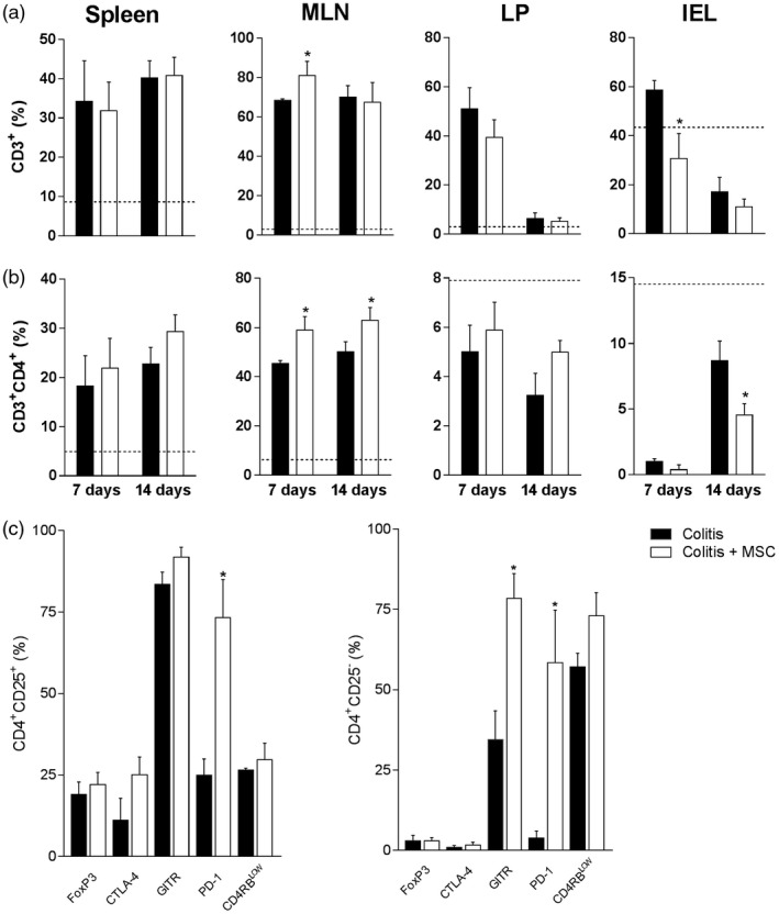
Figure 3.

Mesenchymal stromal cells (MSC) treatment modifies inflammatory infiltrate in the gut mucosa and draining lymphoid organs. Leukocytes from the spleen, mesenteric lymph nodes (MLN), colon lamina propria (LP) and intraepithelial (IEL) compartments of colitis and MSC‐treated mice were isolated and stained with fluorochrome‐conjugated antibodies for flow cytometry on days 7 or 14. After sample acquisition, the frequency of CD3+ T cells (a), CD3+CD4+ (b) or regulatory T cells in the spleen (c, day 14) was determined after analysis by FlowJo software. Results are representative of two independent experiments, with five animals/group, at each time‐point evaluated. Dashed line: control animals treated with ethanol [2,4,6‐trinitrobenzene sulfonic acid (TNBS) (TNBS) vehicle] at day14. (*) Symbols represent significant differences (P < 0·05) from the respective colitis groups.
