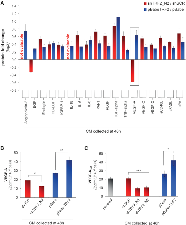
Figure 1.
TRF2 regulates the amount of VEGF-A in the secretome of tumor cells. (A) Luminex/XMAP multiplexed analysis of CMs derived from TRF2-compromised (shTRF2) or -overexpressing (pBabe-TRF2) HCT116 cells and their control counterparts (shScramble/pBabe). CMs were collected 48 h after cell starvation and the expression levels of a panel of secreted chemokines and growth factors involved in angiogenesis were quantified. For each analyte, results are expressed as log2 fold change of protein levels in silenced/overexpressing cells over their controls. (B) Detailed analysis of VEGF-A concentration from A. Histograms show the mean values (±SD) of a single experiment performed in triplicate (*P < 0.1; **P < 0.01; ***P < 0.001; Student's t-test). (C) Concentration of VEGF-A was evaluated by ELISA in the CM of HCT116 silenced (shTRF2_N1 and shTRF2_N2) or overexpressing (pBabe-TRF2) TRF2, collected 48 h after serum-starvation. As control, the amount of VEGF-A was assayed in the CM of HCT116 cells not infected (parental) or infected with viral particles delivering control vectors (shSCR or pBabe). Results were normalized to cell number. Histograms show the mean (±SD) of at least three independent experiments performed in triplicate (*P < 0.1, **P < 0.01, ***P < 0.001; Student's t-test).

