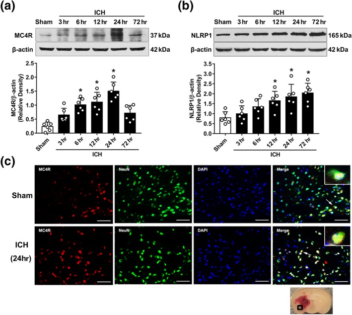
Figure 2.

Endogenous expression of MC4 receptor and NLRP1 inflammasomes after ICH. (a) Representative western blot bands and quantitative analyses of the time‐dependent expression of MC4 receptor (MC4R) from the ipsilateral hemisphere after ICH. (b) Representative western blot bands and quantitative analyses of NLRP1 inflammasome time‐dependent expression from the ipsilateral hemisphere after ICH. Data shown are individual values with mean ± SD; n = 6. *P < 0.05, significantly different from sham; one‐way ANOVA, Tukey's test. (c) Representative microphotograph of double immunofluorescence staining showed the co‐localization of MC4 receptor (MC4R, red) with neurons (NeuN, green) in the perihaematomal area in sham and ICH (24 hr) animals. Nuclei were stained with DAPI (blue). Scale bar = 50 μm, n = 2
