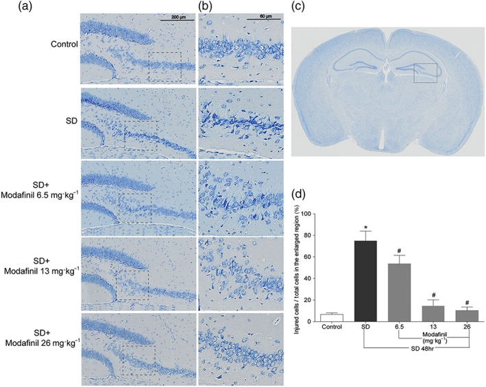
Figure 3.

The effect of modafinil on hippocampal neuronal morphology of mice after sleep deprivation (SD) for 48 hr. (a) Nissl staining of the dentate gyrus and CA3 of hippocampus. (b) The enlarged regions within respective rectangular boxes of (a). (c) An illustration of mouse brain section. The region in the rectangular box was observed for Nissl staining. (d) The percentage of injured cells in the enlarged CA3 region of hippocampus. All data presented are means ± SEM; N = 5 mice per group. *P < 0.05; significantly different from control. # P < 0.05, significantly different from SD group
