
Figure 7.
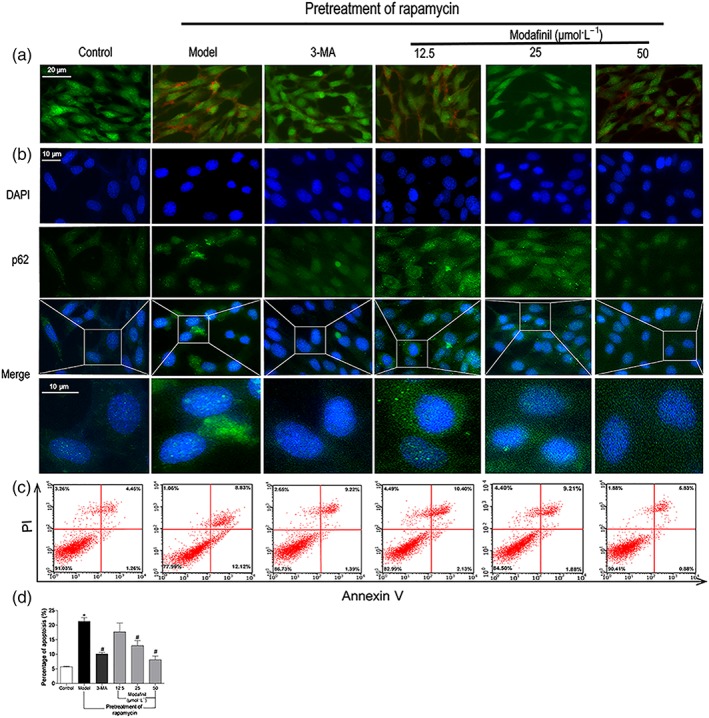
The effect of modafinil on autophagy and apoptosis of HT‐22 cells. (a) Acridine orange staining showed that modafinil could reduce acidic vesicular organelles in bright red colour in HT‐22 cells induced by rapamycin (Model). (b) Immunostaining of p62 exposed that modafinil could decrease p62 expression in HT‐22 cells induced by rapamycin. (c) AnnexinV/propidium iodide staining showed that modafinil could reduce the percentage of apoptotic HT‐22 cells induced by rapamycin. The right upper and lower quadrants of flow cytometry plot depicted the late apoptosis and early apoptosis respectively. (d) The total percentage of the early and late apoptotic cells. Generally, the total percentage of the early and late apoptotic cells was used to represent total apoptotic cells. All data presented are means ± SEM; N=3. *P < 0.05, significantly different from control; # P < 0.05, significantly different from Model group