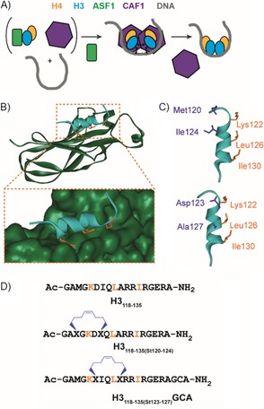
Figure 1.

ASF1 as a target for constrained peptides. A) Schematic illustration of the role of ASF1 (green) in displacing CAF‐1 (purple) through the recognition of histone H3 (cyan) and H4 (yellow) so as to facilitate nucleosome formation. B) Structure of the histone H3(118–135) (cyan)–ASF1A(1–156) (dark green) interaction as determined by NMR spectroscopy (PDB ID: 2IIJ)[45]—the histone side chains located on one face that are perceived to be important for binding are shown as orange sticks. C) The key H3 helix (cyan), key side chains (orange) and residues at i, i+4 positions considered suitable for introduction of a constraint (purple) are highlighted. D) Sequences of the peptides used in this study with the positions of the hydrocarbon constraints.
