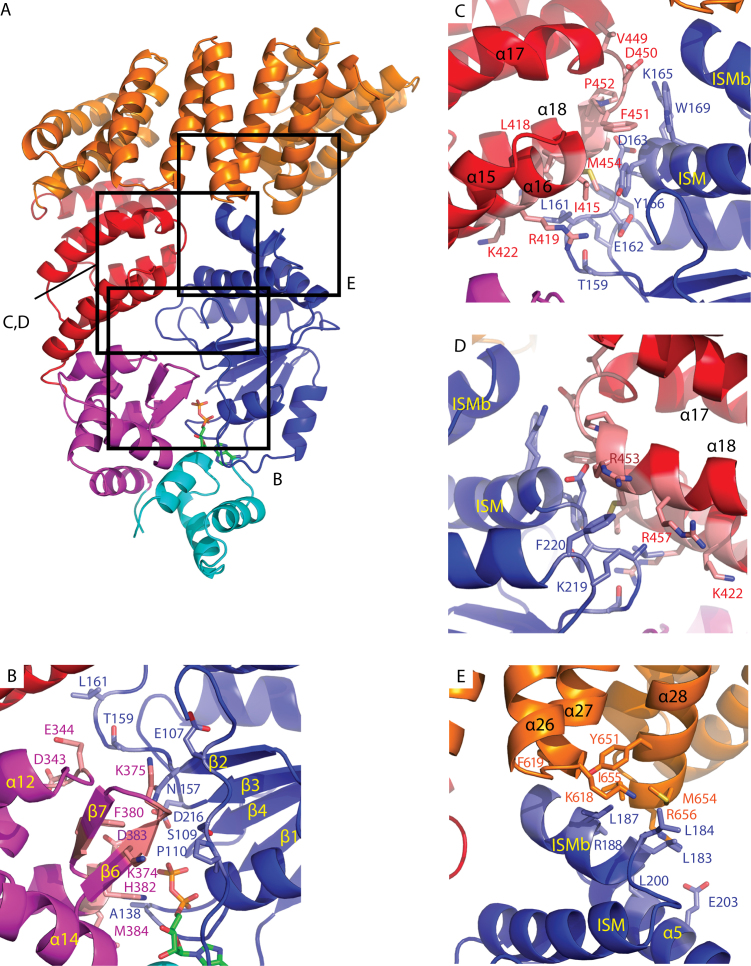
Figure 3.
NBD interactions with non-adjacent domains in the PH0952 crystal structure. The protein is depicted in cartoon representation (PDB code: 6MFV). Highlighted residues are shown in sticks in the zoomed panels. (A) Overall view of PH0952ΔN. Squares highlight regions enlarged in the other panels. (B) NBD–WHD interface, (C) NBD–arm interface, (D) NBD–arm interface, rear view and (E) NBD–sensor.

