
Figure 1.
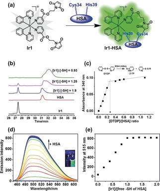
a) The conjugation of Ir1 to HSA. b) Reaction of Ir1 (30 μm) with various concentrations of HSA (0, 60, 90, and 120 μm, concentrations of Cys34 free thiol group were 0, 16.2, 24.3, 32.4 μm, respectively) studied by RP‐HPLC (UV detection at 280 nm). c) Variation of absorbance at 340 nm at various HSA:2,2′‐DTDP ratios ([HSA]=80 μm; [DTDP]=0–110 μm) and determination of the thiol content of HSA. d) Emission spectra of Ir1 (4 μm) in the presence of increasing concentrations of HSA (0–30 μm, [Cys34 free thiol] 0–8.1 μm), in PBS (pH 7.4), reaction time: 20 min at each concentration, λ ex=405 nm; Inset: photos of Ir1 (1) and Ir1‐HSA (2) under UVA irradiation. e) Dependence of the phosphorescence intensity of Ir1 at 515 nm on the concentration of free thiol of HSA, as ratio [Ir1]/[free thiol].