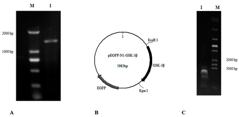
Figure 1.
The construction of pEGFP-N1-GSK-3β. (A) The ORF of GSK-3β was cloned according to primers GSK-3β-F1 and GSK-3β-R1. Lane 1, DNA marker; Lane 2, GSK-3β. (B) The map of pEGFP-N1-GSK-3β. (C) The connected fragment of GSK-3β and pEGFP-N1 was amplified using PCR. Lane 1, the connected fragment of GSK-3β and pEGFP-N1; Lane 2, DNA marker.

