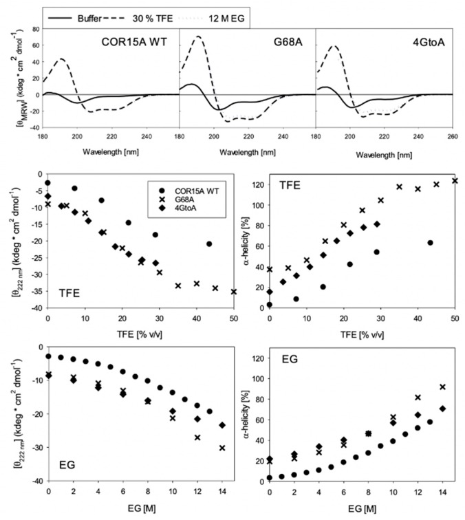
Figure 7.
Far-ultraviolet (UV) circular dichroism (CD) spectra in buffer, 20 (v/v) % TFE and 12 M Ethylene Glycol for COR15A WT, G68A and 4GtoA (A). The mutants have more α-helical spectra than the WT in buffer alone, and in the presence of high concentrations of both co-solvents. Coil-helix transitions of COR15A WT and both mutants in TFE (B) and EG (C), specified by θMRW at 222 nm (left panels) and derived α-helix ratios (right panels).

