Figure 5.
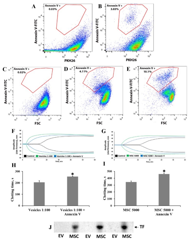
Analysis of phosphatidylserine and tissue factor (TF) expression on MSC and EV surface and the role of phosphatidylserine in procoagulation effects. Elucidation of the mechanisms of MSC- or EV-induced coagulation. MSC-derived EVs were analyzed by flow cytometry with (A) PKH26, a lipid-staining dye, or (B) a combination of PKH26 and fluorescein isothiocyanate (FITC)-conjugated annexin V. Flow cytometry analysis of phosphatidylserine exposure on the MSC surface using annexin V-FITC: (C) unstained MSCs, (D) annexin V-FITC stained native MSCs, and (E) annexin V-FITC stained MSCs after apoptosis induction. Effect of phosphatidylserine masking on blood coagulation: preincubation of (F,H) EVs and (G,I) MSCs with annexin V to shield phosphatidylserine decreased blood coagulation, as shown by thromboelastometry. (F,G) Representative thromboelastograms and (H,I) calculated clotting time assayed with the NATEM test. MSCs and EVs were preincubated with annexin V (see Methods), then resuspended in 1 mL of freshly obtained blood and analyzed by the NATEM test. Experiments used blood from three donors and each blood sample was incubated with MSCs from different umbilical cord samples. * p < 0.05, Student’s t-test. (J) Detection of TF on Western blots of MSC and EV samples.

