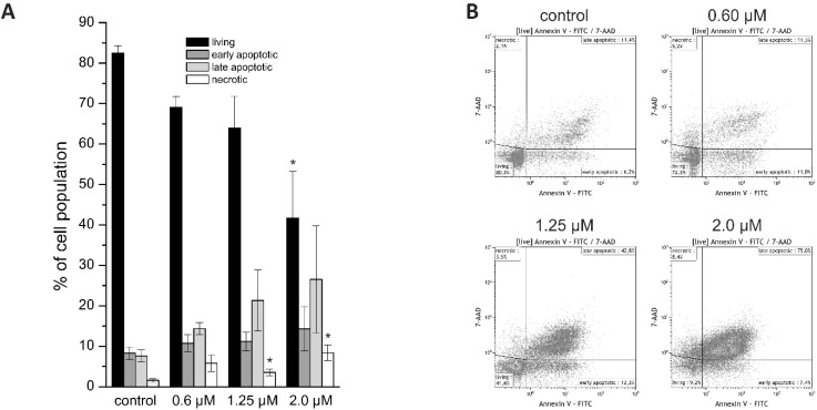
Figure 2.
(A) Annexin V/7-AAD staining of HuCCt-1 cells after 24 h incubation time with 0 (control), 0.6, 1.25, or 2.0 µM napabucasin. (B) Exemplary Annexin V/7-AAD staining scatter plots. Data are presented as mean value ± SEM related of at least three individual biological replicates * indicates significant (p < 0.05) and ** highly significant (p < 0.01) results. FITC: fluorescein isothiocyanate.

