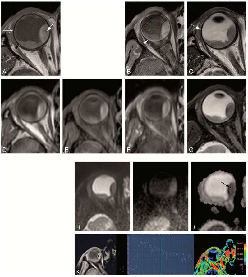
Figure 11.
(A–K) Clinical protocol with anatomical and functional sequences in a patient with a uveal melanoma of the right eye (arrow). (A) MS 2 mm T1. (B) MS 2 mm enhanced T1 with fat signal suppression. (C) MS 2 mm T2. (D) 3D TSE 1 mm T1. (E) 3D TSE 1 mm T1 with fat signal suppression. (F) 3D TSE 1 mm enhanced T1 with fat signal suppression. (G) 3D TSE 0.8 mm T2 with fat signal suppression. (H–J) TSE DWI, with b values of 0 (H), 800 (I) and ADC (J). Restricted diffusion in the uveal melanoma (black arrow). (K) DCE with a good quality, few motion artefacts, showing a wash-out TIC pattern. Notice that on T2 WI the globe wall appears as a hypointense line (dashed arrow) and the different layers cannot be separated. On T1 the outer hypointense line corresponds to the sclera (open arrow), while the inner hyperintense layer corresponds to both the choroid and retina (black arrowhead). After contrast only the choroid and retina enhance (arrowhead). Anteriorly the ciliary body and iris can also be identified. The aqueous humor and vitreous body have a signal intensity similar to water. On the contrary, the lens is hypointense on T2 and slightly hyperintense on T1.

