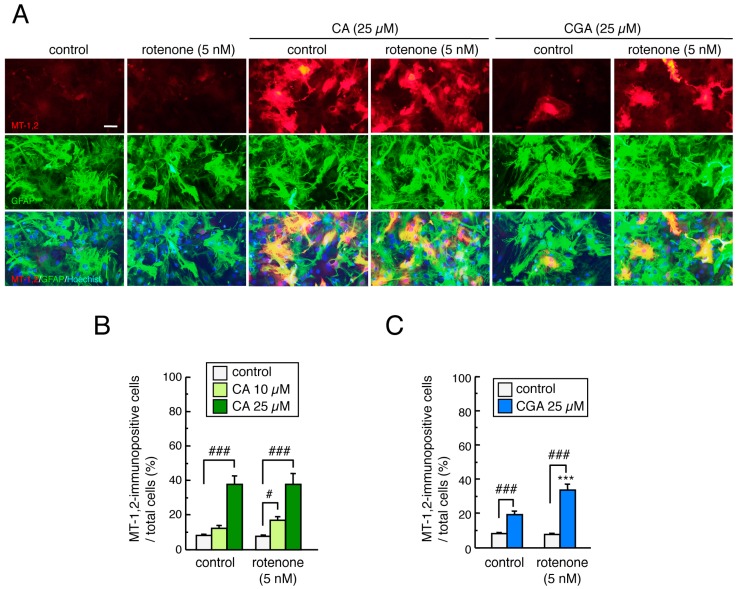
Figure 6.
Effects of treatment with CA or CGA followed by rotenone exposure on MT-1,2 expression in mesencephalic astrocyte cultures. Astrocyte cultures were pretreated with CA (10 or 25 µM) or CGA (25 µM) for 24 h and co-treated with rotenone (1–5 nM) for a further 48 h. (A) Representative photomicrographs of MT-1,2 and GFAP double immunostaining in astrocyte cultures. Red: MT-1,2-positive signals. Green: GFAP-positive astrocytes. Blue: nuclear staining with Hoechst 33342. Scale bar = 50 µm. (B,C) Quantitation of MT-1,2-positive signals in astrocyte cultures after treatment with rotenone and CA (B) or CGA (C). Each value is the mean ± SEM (n = 8–13) expressed as the percentage of the MT-1,2-immunopositive astrocytes in the total cell population. *** p < 0.001 vs. each control group, # p < 0.05, ### p < 0.001 between the two indicated groups.

