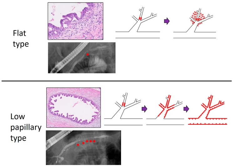
Figure 2.
Comparison of flat and low papillary type of PC in situ (PCIS). Histologically, the case with flat type PCIS seemed to invade with little intraductal spread, whereas the case with low papillary type PCIS tended to change to invasive PC after spreading intraductally. Red dots in the ERCP image indicate the position of PCIS. Purple bar: tumor progression. Red line with triangles: tumor position. Magnification of histological slides: 10×. This study is approved by the review board of research ethics committee in Onomichi General Hospital (approval No: OJH-201862, approval date: 16 January 2019).

