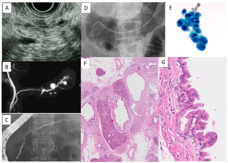
Figure 4.
A case with PCIS (58 years old, female). (A): Endoscopic ultrasonography (EUS) finding in the pancreas body, (B): magnetic resonance cholangiopancreatography (MRCP), (C): ERCP, (D): ENPD, (E): Cytologic findings using pancreatic juice, (F): Histological findings. The red arrow indicates the location of PCIS in MPD. Magnification: 4×. (G): The enlarged view of PCIS in the main pancreatic duct (MPD). Magnification: 40×. This study is approved by the review board of research ethics committee in Onomichi General Hospital (approval No: OJH-201862, approval date: 16 January 2019).

