Figure 1.
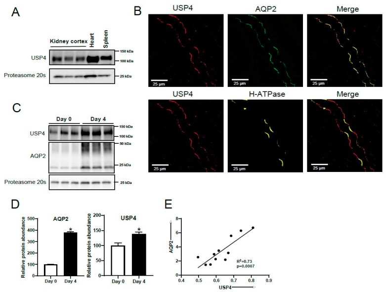
USP4 is expressed in the collecting duct in vivo and its abundance is regulated by vasopressin in vitro. (A) Immunoblotting using a USP4-specific antibody demonstrated USP4 to be abundantly expressed in mouse kidney cortex. Each lane represents a sample from an individual mouse, and heart and spleen tissues are positive controls. (B) Confocal microscopy images of mouse kidney sections immunolabeled with USP4, the principal cell marker AQP2, and the H+ATPase B1 subunit, an intercalated cell marker. AQP2 and USP4 co-localize at the apical plasma membrane of principal cells. (C) Representative immunoblot images of USP4 and AQP2 in mpkCCD14 cells cultured in dDAVP for 4 days. (D) Summarized data relative to day 0 (normalized to proteasome 20s). * indicates p < 0.05 relative to day 0. (E) Linear regression analysis of AQP2 and USP4 protein abundance in mpkCCD14 cells after dDAVP treatment for various periods (0, 24, 48, and 72 h).

