Figure 3.
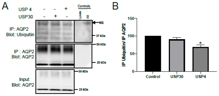
USP4 deubiquitylates AQP2 in vitro. mpkCCD14 cells were grown in the presence of dDAVP for 4 days to induce AQP2 expression. On the day of the experiment, cells were washed twice with DMEM/F12 media and subsequently treated with 25 nM 12-O-tetradecanoylphorbol-13-acetate (TPA) for 15 min at 37 °C. AQP2 was immunoprecipitated (IP) and incubated with either USP4 (100 nM) or USP30 (negative control, 100 nM). (A) Representative immunoblots of ubiquitylated and total AQP2 levels following the deubiquitylation assay. + and − indicates presence or absence of the DUB respectively (B) Summarized data (n = 3, performed on different days) relative to control. * indicates p < 0.05 relative to control conditions. Breaks in the images are to show the immunoprecipitation controls (antibody (AB) alone or lysate alone) next to the samples. NS = non-specific band.

