Figure 2.
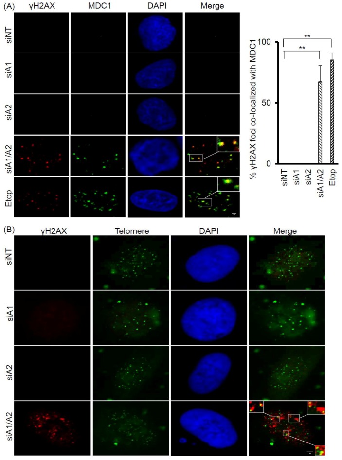
Co-localization of γH2AX with MDC1 and telomere DNA in A549 cells depleted of hnRNP A1/A2. (A) A549 cells were transfected with siRNA targeting hnRNP A1 (siA1), hnRNP A2 (siA2), both hnRNP A1 and A2 (siA1/A2), or with a non-targeting sequence (siNT) for 72 h. Cells were fixed and immunostained for γH2AX (red) and MDC1 (green), and nuclei were counterstained with DAPI (blue). A549 cells treated with 50 μM etoposide (Etop) for 12 h served as a positive control. The percentage of γH2AX foci that co-localized with MDC1 (see marked squares for examples) was determined from 50 γH2AX-positive nuclei; data from 3 experiments are summarized in the right panel. ** p < 0.01 versus siNT control; The bar equals 2 μm.(B) Cells were stained for γH2AX (red) and then for telomeric DNA using FISH with fluorescein isothiocyanate (FITC) -conjugated oligonucleotides (green). γH2AX foci that colocalized with telomere DNA are illustrated in the boxed regions. The scale bar equals 2 μm.

