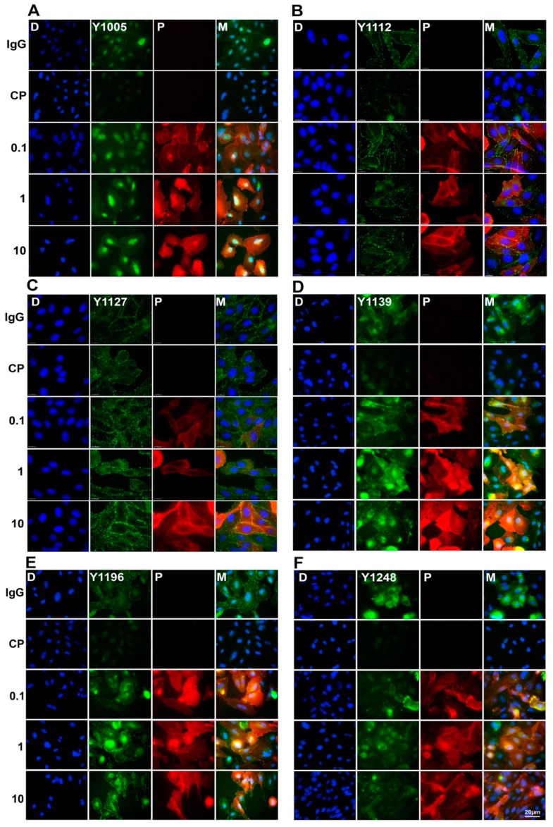
Figure 4.
Double-immunofluorescence staining of pHER2 and pertuzumab in pertuzumab treated CHO-K6 cells. CHO-K6 cells were treated with 0.1, 1 and 10 μg/mL pertuzumab for 60 min then pHER2 at (A) pY1005 (B) pY1112, (C) pY1127, (D) pY1139, (E) pY1196, and (F) pY1248 HER2 (all green) and pertuzumab (red) were stained. Ten μg/mL human IgG and 20 μM CP-724714 (CP) were used as mock and HER2 positive controls, respectively.

