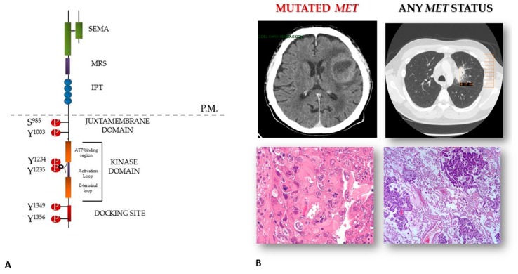
Figure 1.
MET activation in brain lesions from non-small cell lung cancer (NSCLC). Panel (A) structure of the MET receptor; panel (B) brain metastases and the matched primary lung mass. The lesions feature similar histo-morphology, but different MET status, with MET mutated cells being only detected in the secondary lesion.

