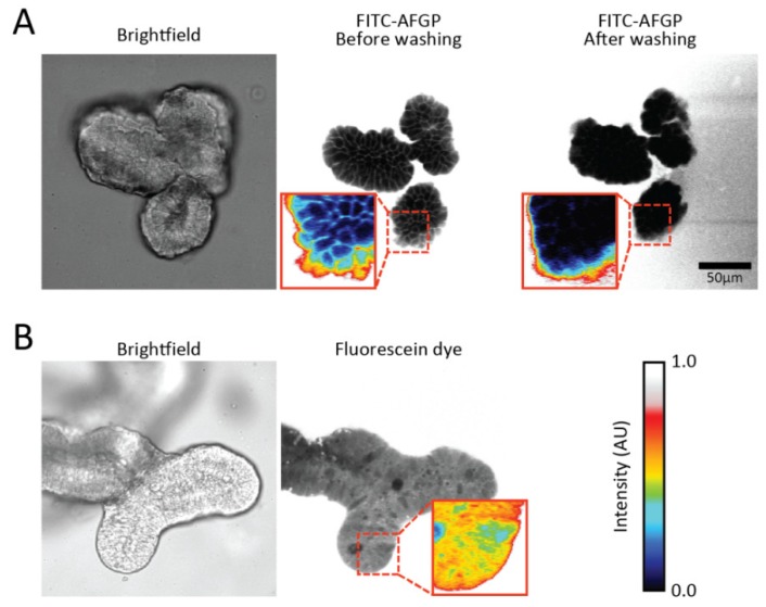
Figure 3.
Fluorescence images of organoids in the presence of FITC-labeled AFGP. (a) Brightfield and fluorescence images of an organoid with FITC-AFGP in the medium reveal that fluorescence is observed on the cell outlines. Washing of the imaging wells with buffer eliminated the fluorescence pattern; (b) Brightfield and fluorescence images of an organoid with fluorescein diacetate in the medium show that fluorescence is present in the cytoplasm of most cells. Pixel intensities are colored in insets to highlight the observed fluorescence patterns. Colors represent the same intensities in all images.

