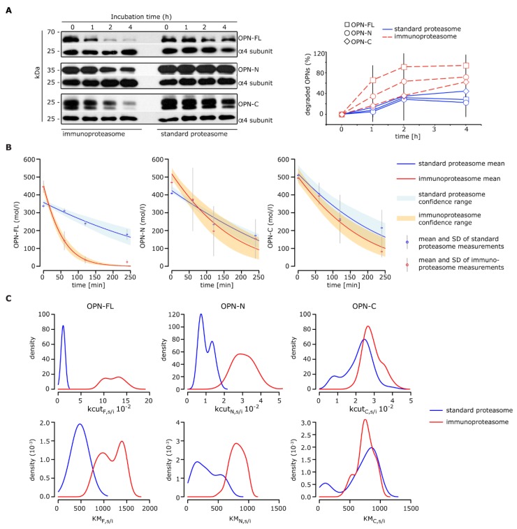
Figure 2.
Processing of OPNs by standard- and immuno-proteasomes. (A) Representative Western Blot staining of the degradation kinetics of OPNs (OPN-FL, OPN-N, and OPN-C) by 20S standard- and immuno-proteasomes (left panel; the proteasome α4 subunit is used as control marker); the corresponding relative OPN’s degradation is also depicted (right panel; mean and SD of two to three independent experiments measured in duplicate are shown). (B–C) Inference of kinetic parameters determining the OPNs degradation dynamics by standard- and immuno-proteasomes. Shown are the model fits based on simple Michaelis-Menten kinetics (B) and the inferred marginal posterior parameter distributions for kcut (maximal reaction velocity) and KM (Michaelis-Menten constant) using Bayesian inference via the Metropolis-Hastings algorithm (C).

