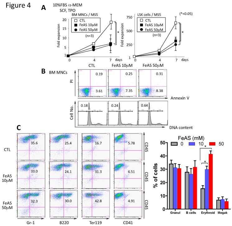
Figure 4.
FeAS inhibits the growth of murine BM cells and modulates lineage distribution after culture with MS5 cells. (A) BM MNCs (1 × 10⁴ cells) or LSK cells (100 cells/well) were co-cultured with MS5 cells under the indicated conditions for 7 days. Cell proliferation was determined by counting the total viable cells using the ATP assay. The data shown are representative of three independent experiments (* p < 0.05). (B) The proportion of apoptotic cells was determined with annexinV/PI-staining. The DNA content of cells cultured for 7 days was examined by PI staining. The proportion of cells in annexin V+ and the sub-G1 fraction is indicated, respectively. (C) After 7 days of culture, specific antibodies were used to characterize the surface phenotype of cultured LSK cells as indicated. Representative fluorescence-activated cell sorting data from one experiment are shown (left panel) and summarized data (mean ± SD) from three independent experiments is shown in the right panel (* p < 0.05) (Gr-1+ granulocytes, B220 B cells, Ter119 erythroid cells and CD41 megakaryocytes).

