Figure 5.
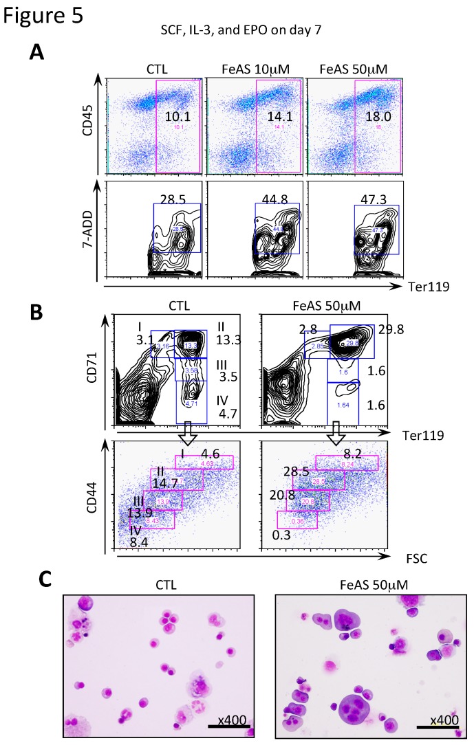
FeAS interferes with the development of erythroid cells with severe morphologic abnormalities. (A) Flk-1+ cells were induced to develop into erythroid cells using the OP9 system as described in Materials and Methods. After 7 days of culture with the indicated cytokine cocktail in the presence or absence of FeAS, the expression of CD45 and Ter119 on the cultured cells was examined by flow cytometry analysis. Apoptotic cells in the erythroid lineage were detected as Ter119+7-AAD+ cells, and the percentage of these cells is indicated. (B) Erythroblasts at different maturation stages were identified by double staining with FITC-conjugated anti-TER119 and APC-conjugated anti-CD44 Abs. Plots of CD44 vs. forward scatter (FSC) for TRE119-positive cells are shown. After cells were cultured as described above, the expression of CD71 and Ter119 was examined by flow cytometry analysis. The percentages of cells in fractions I to IV are indicated. I (Ter119lowCD71+) corresponds to ~CFU-E; II (Ter119+CD71+) to proerythroblasts; III (Ter119+CD71 low) to erythroblasts; IV (Ter119+CD71−) to erythrocytes. (C) After 11.5 days of culture in the absence or presence of 50 μM FeAS, cultured cells were subjected to morphological analysis.

