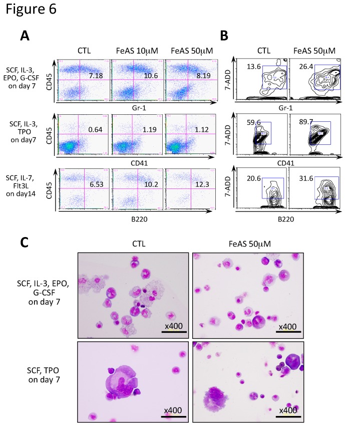
Figure 6.
FeAS shows little influence on the development of myeloid cells, megakaryocytes, and B lymphocytes. (A,B) Myeloid cells, megakaryocytes, and B lymphocytes were induced to develop under the indicated conditions using the OP9 system, and the respective lineage markers, Gr-1, CD41 and B220, were examined by flow cytometry analysis. Reactivity to 7-AAD was also analyzed by flow cytometry. The percentage of each fraction is indicated. (C) The morphology of granulocytes and megakaryocytes treated with FeAS obtained by the above-described culture conditions were analyzed.

