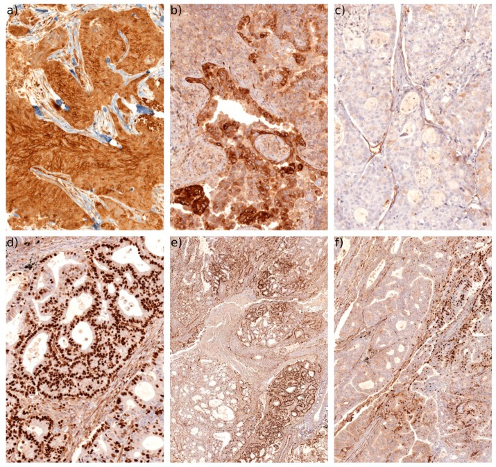
Figure 5.
IHC of Cx43. (a) Typical example in LUSC demonstrating intense Cx43 staining with a predominantly membranous and cytoplamsic pattern (20×). (b) Typical example in LUAD of a patchy high Cx43 expression with a predominantly cytoplasmic expression pattern (20×). (c) Example of a LUAD with very low or negative Cx43 expression (20×). Note that stromal cells such as endothelial cells are still positively stained. (d) Typical example of high levels of nuclear Cx43 expression in LUAD (20×). Note that some non-tumour cells are also sometimes positive for nuclear Cx43. (e) Low magnification overview (4×) of a LUAD with significant areas of nuclear Cx43 expression. (f) Area of the same tumour (10×) where nuclear Cx43 can be observed together with areas either negative for Cx43 or with low cytoplasmic levels.

