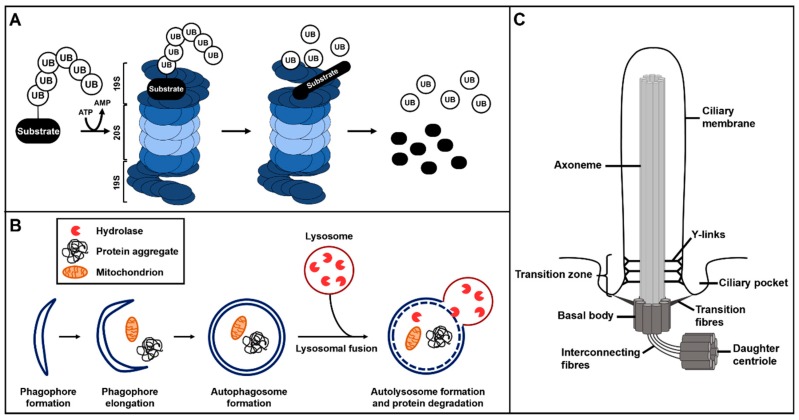
Figure 1.
Overview of ubiquitin–proteasome system (UPS) protein degradation, autophagy and primary cilium structure. (A) The ubiquitin–proteasome system. The ubiquitinated substrate is recognised by the 19S regulatory subunit of the 26S proteasome and gets degraded or proteolytically processed by the 20S subunit of the 26S proteasome. (B) Autophagy starts with the formation of phagophores which subsequently elongate to finally develop into autophagosomes. During autophagosome formation, target proteins and structures become enclosed in the autophagosomes. These autophagosomes fuse with lysosomes and the hydrolases of the lysosomes degrade the target proteins and structures. (C) Primary cilia consist of a microtubule scaffold called axoneme. The axoneme is surrounded by the ciliary membrane and grows out of the basal body. The basal body is a modified mother centriole that is connected to the daughter centriole by interconnecting fibres. The basal body is attached to the ciliary membrane in the region of the ciliary pocket via transition fibres. The transition zone, with its Y-links, is located at the proximal part of the axoneme.

