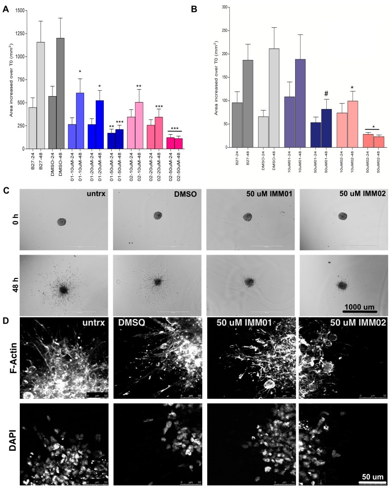
Figure 3.
mDia agonists halt high-grade glioma (HGG) patient neuro-sphere invasion. Neuro-spheres were formed and embedded in matrigel. Neuro-sphere areas were measured at embedding (T0), at 24 h and 48 h invasion. (A) Change in neuro-sphere invasion graphed as the change in area over T0 for Pat9. N > 12 neuro-spheres/condition over three replicate experiments. p values are relative to the corresponding time point in B27-treated control invasion assays where * p < 0.05; ** p < 0.02; *** p < 0.006. (B) Change in neuro-sphere invasion graphed as the change in area T0 for Pat8. N > 12 neuro-spheres/condition over three replicate experiments. p values are relative to the corresponding time point in B27-treated control invasion assays where * p < 0.0001; # p < 0.003. (C) Representative images of Pat9 neuro-sphere invasion at 0 and 48 h. Scale bars = 1000 μm. (D) Confocal images of fixed Pat9 48 h invasion assay stained for phalloidin and DAPI and showing the neuro-sphere edges. Scale bars = 50 μm.

