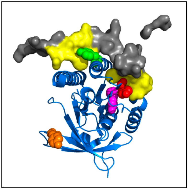
Figure 10.
Modeling of tyrosine residues of RAD51 involved in the RAD51-BRC4 interface. Using the PyMOL software and the structure of the C-terminal part of RAD51 with BRC4 [25], tyrosines (Y159, magenta; Y191, red; Y205, green; Y315, orange) were indicated. The BRC4 peptide [26] and its high interaction areas with RAD51 are also represented (BRC4, grey, surface; high interaction with RAD51, yellow, surface).

