Figure 2.
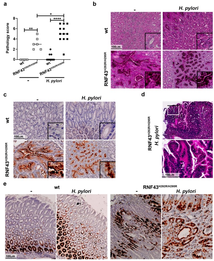
H. pylori enhances gastric pathology of RNF43H292R/H295R mice. (a) Pathology score. The presence of atrophy, metaplasia, hyperplasia and reactive changes was assessed in antrum and corpus after 6-month H. pylori infection. (b) Representative images of periodic acid Schiff (PAS) stained gastric sections. (c) MUC2 expression detected in gastric tissue sections by immunohistochemistry. (d) Representative picture and higher magnification image of hyperplasia in the stomach of an infected RNF43H292R/H295R mouse. * p ≤ 0.05; ** p ≤ 0.01; **** p ≤ 0.0001. Mann-Whitney Test (pairwise comparisons). (e) Ki67 staining denoting cellular proliferation in the stomach.

