Figure 1.
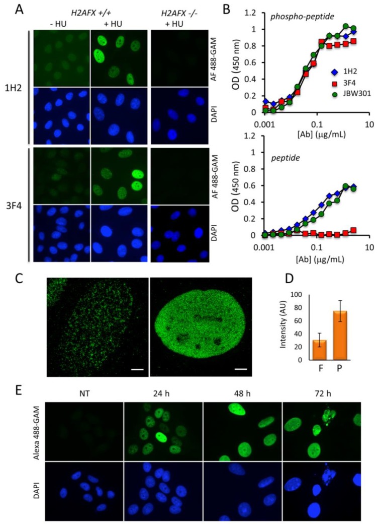
Detection of HU-induced γ-H2AX in HeLa cells with the newly-generated antibodies. (A) After treatment with HU for 24 h, either wild type or H2AFX−/− HeLa cells were subjected to analysis by immunofluorescence with a conventional microscope (Materials and Methods). The binding of mAb 1H2 or mAb 3F4 was revealed with Alexa Fluor 488-labeled goat anti-mouse immunoglobulins (green). The nuclei were counterstained with DAPI (blue). Magnification: 630×. (B) Analysis of the binding specificity of mAbs 1H2, 3F4 and JBW301 by ELISA. After incubation in the presence of either the phosphorylated (phospho-peptide) or the nonphosphorylated (peptide) peptides corresponding to the C-terminus of H2AX coated on plate, bound mAbs were revealed with HRP-labeled secondary anti-mouse globulins. The curves summarize the data obtained in 2 independent experiments. The color code is indicated. (C) The HeLa cells stained with mAb 3F4 shown in A were analyzed by 3D-structural illumination super resolution microscopy (3D-SIM). Typical cells with either foci (left) or nuclear-wide γ-H2AX staining (right) are shown. Scale bar: 4 μm. (D) The average nuclear fluorescence intensity of a minimum of 200 HeLa cells harboring either γ-H2AX foci (F) or pan-nuclear γ-H2AX (P) after staining with mAb 3F4 as in A is represented. (E) HeLa cells were incubated with HU and fixed 24 h, 48 h, and 72 h post-treatment. γ-H2AX levels were analyzed as in (A).

