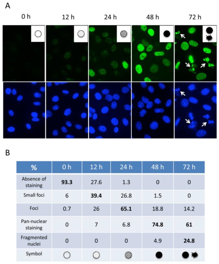
Figure 3.
Dynamics of γ-H2AX formation in HeLa cells following treatment with HU. (A) Typical patterns of γ-H2AX staining observed during the analysis with mAb 3F4 by immunofluorescence are shown and represented as symbols (insets). Cells with fragmented nuclei at 72 h post-treatment are indicated (white arrows). Magnification: 630×. The assignment of the symbols that correspond to the major characteristic γ-H2AX patterns observed in (A) is shown (B). Up to 160 nuclei recorded from three independent experiments at each time point were analyzed to calculate the percentages.

