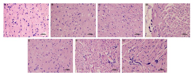
Figure 4.
H&E staining demonstrating the morphology of myocardial cells in each group. Magnification, ×200. (A) Sham, (B) sham + NaHS, (C) sham + LY, (D) CLP, (E) CLP + NaHS, (F) CLP + LY and (G) CLP + NaHS + LY group. H&E, hematoxylin and eosin; NaHS, sodium hydrosulfide; CLP, cecal ligation and puncture; LY, LY294002.

