
Figure 1.
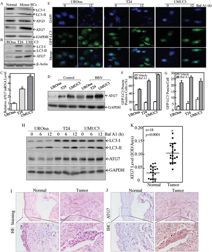
ATG7 was overexpressed in mouse invasive BCs, human invasive BC cells, and tissues. A) Western Blot assay was performed to detect the conversion of LC3 from LC3‐I to LC3‐II, ATG3 and ATG7 expression in mouse invasive BCs (n = 5). B) Western Blot was used to determine the conversion of LC3 from LC3‐I to LC3‐II and ATG7 protein expression, β‐Actin was used as a protein loading control. C) Real‐time PCR was performed to detect ATG7 mRNA expression, and the asterisk (*) indicates a significant increase from normal UROtsa cells (p < 0.05). D) UROtsa, T24, and UMUC3 cells were seeded into six‐well plates and the cells were then treated with or without 400 × 10−6 m of BBN for 24 h. The cell extracts were subjected to Western Blot for the determination of protein expression as indicated. GAPDH was used as a protein loading control. E) The GFP‐LC3 construct was stably transfected into UROtsa, T24, and UMUC3 cells, and then treated with 5 × 10⁻⁹ m Baf A1 for 12h. LC3 puncta formation was observed and images were captured using fluorescence microscopy. F,G) Percentage of GFP‐LC3 puncta cells (F) and the number of puncta per positive cell (G) were calculated. The asterisk (*) indicates a significant increase as comparison to UROtsa cells treated with Baf A1 (p < 0.05). H) Western Blot was performed to determine autophagy flux and ATG7 expression in presence of 5 × 10⁻⁹ m of Baf A1. I,J) Hematoxylin‐eosin (HE) and IHC staining were performed to evaluate morphology and ATG7 expression in 18 paired human BC tissues and their adjacent normal bladder tissues. The IHC images were captured using the AxioVision Rel.4.6 computerized image system. K) The ATG7 protein expression levels were analyzed by calculating the integrated IOD/area using Image‐Pro Plus version 6.0. Three independent experiments were performed, the Student's t‐test was utilized to determine the p‐value; and the asterisk (*) indicates a significant increase from the adjacent normal bladder tissues (*p < 0.05).