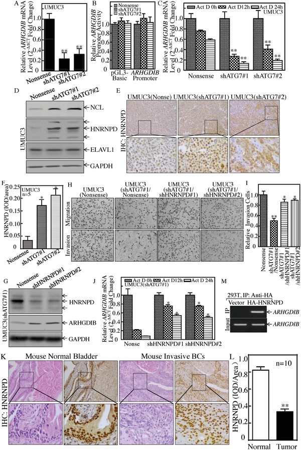
Figure 6.

HNRNPD was an ATG7 downstream effector and inhibited ARHGDIB mRNA stability and BC cell invasion. A) Real‐time PCR was used to determine ARHGDIB mRNA expression, and β‐Actin was used as an internal control, **p < 0.05. B) Human ARHGDIB promoter‐driven luciferase activity was evaluated in the indicated cells, p > 0.05. C) ARHGDIB mRNA stability was detected in the presence of Act D by using real‐time PCR, **p < 0.05. D) The indicated cell extracts were subjected to Western Blot for determination of NCL, ELAVL1, and HNRNPD protein expression. E,F) IHC staining was performed to detect HNRNPD expression in the tumor tissues obtained from the nude mice injected with UMUC3(shATG7#1), UMUC3(shATG7#2), or UMUC3(Nonsense) cells, *p < 0.05. G–I) HNRNPD knockdown constructs were stably transfected into UMUC3(shATG7#1). The HNRNPD knockdown efficiency was evaluated by Western Blotting (G). The stable transfectants were used to determine their invasion abilities in comparison to nonsense control transfectants (H and I). Student's t‐test was utilized to determine the p‐value, the double asterisk (**) indicates a significant decrease in comparison to scramble vector transfectants (**p < 0.05), while the asterisk (*) indicates a significant increase in comparison to UMUC3(shATG7#1/Nonsense) transfectants (*p < 0.05) (I). J) ARHGDIB mRNA stability was evaluated by real‐time PCR in the presence of Act D in UMUC3(shATG7#1/shHNRNPD) cells and its nonsense control, *p < 0.05. K,L) IHC staining was performed to evaluate HNRNPD expression in mouse highly invasive BCs, **p < 0.05. M) HA‐HNRNPD construct was transfected into 293T cells and HA‐HNRNPD protein was pulled down with anti‐HA beads. The mRNAs bound to HNRNPD protein were used to carry out RT‐PCR for determination of ARHGDIB mRNA expression.
