Figure 1.

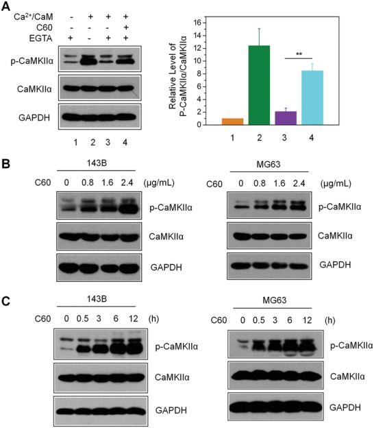
Nano‐C60‐induced autonomous CaMKIIα activity in OS cells. A) T286 autophosphorylation assay for CaMKIIα in 143B cell lysates, as detected by anti‐CaMKII and phospho‐CaMKII antibodies. The right panel shows the level of p‐CaMKIIα relative to total CaMKIIα, with the value for control (without Ca2+/CaM and nano‐C60) set at 1. Mean ± SEM, n = 3. **P < 0.01. B) Dose‐dependent CaMKIIα‐T286 autophosphorylation level in 143B and MG63 cells treated with nano‐C60 for 12 h. C) Time course of CaMKIIα‐T286 autophosphorylation levels in 143B and MG63 cells treated with 2.4 µg mL−1 nano‐C60.
