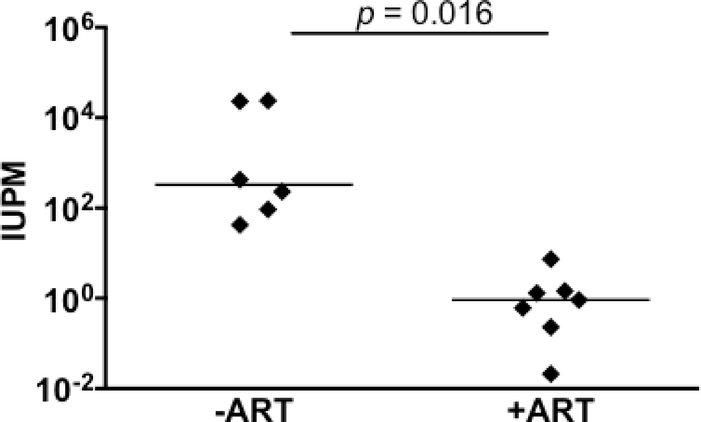
Figure 3.
Quantitation of latently infected brain macrophages in ART-treated macaques by MΦ-QVOA. Quantitation of infected brain macrophages from ART-treated macaques (85). Comparison between the numbers of SIV-infected brain macrophages isolated from animals that were not given ART (-ART) and the numbers isolated from animals that were treated with ART and with viral suppression <10 copies SIV RNA/ml plasma. The horizontal black line represents the median IUPM values. The MΦ QVOA results from SIV-infected animals with and without ART have been reported (85, 94). Significance was determined by Mann-Whitney nonparametric t test; a P of <0.05 was considered significant.

