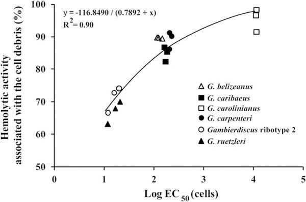
Fig. 4.
Percent of total hemolytic activity associated with the cell debris after the cells had been disrupted using sonication. These data were determined using a representative clone for each of the six species. For each isolate, the % hemolysis was determined when the cells were in log, late log – early stationary and mid-stationary phase (3 data points for each species).

