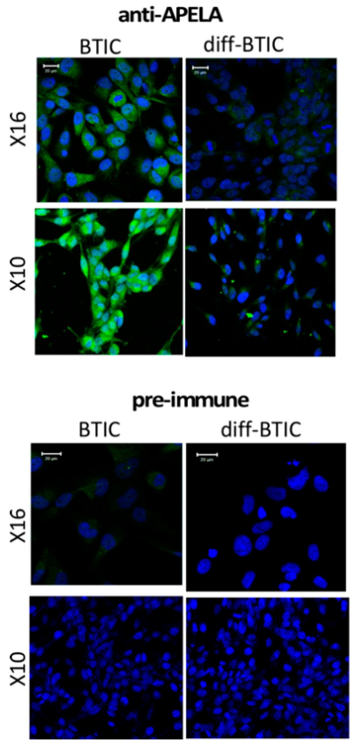
Figure 2.

Immunohistochemistry (IHC) for APELA protein expression in BTICs and differentiated BTICs. IHC was performed on bulk differentiated and X10 and X16 BTICs. Staining was performed with APELA antisera or pre-immune sera and processed for IHC. Nuclei were counterstained with 4’,6-Diamidine-2’-phenylindole dihydrochloride (DAPI). Note, the markedly more intense cytoplasmic staining for APELA in BTICs than in bulk GBM cells, and no detectable staining obtained with pre-immune sera. Representative images are shown. Staining with pre-immune sera showed no IHC staining.
