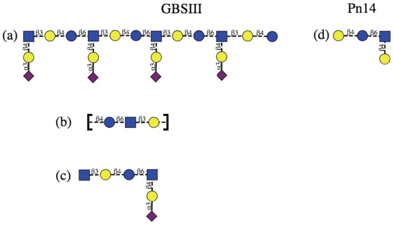
Figure 1.
Schematic representation of the protective epitopes previously identified for GBS PS (left column) and Pn14 PS (right column). GBSIII: (a) 3–4 RU helical conformational epitope postulated for GBSIII PS [10,11]; (b) the linear backbone epitope identified from fragment binding to GBSIII antibodies [14] and (c) a 6-residue epitope identified from a DP2-Fab crystal structure [13]. Pn14: (d) the tetrasaccharide epitope first identifed by Safari et al. [7,14] and then by Kurbatova et al. [15] from antibody studies. Structures are depicted using the ESN symbol set [16] with yellow circle: Gal, blue circle: Glc, blue square: GlcNAc, purple diamond: Neu5Ac.

