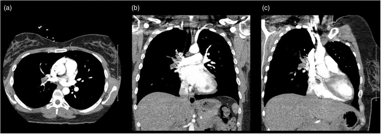
Figure 1.
Computed tomography (CT) scan of the chest with contrast. CT scan of the chest done using pulmonary embolism (PE) protocol that demonstrates extensive PE extending throughout the entire right pulmonary artery with sparing of the left pulmonary artery, seen in (a) axial, (b) coronal, and (c) sagittal views. Panel (c) also demonstrates the right atrial myxoma, which was identified as the source of the PE.

