
Figure 3. Effect of alcohol feeding on mitochondrial total Ca2+ content and mass in freshly isolated hepatocytes.
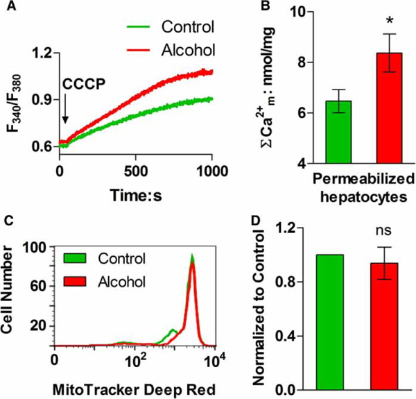
(A) Representative traces of CCCP-induced Ca2+ release in digitonin-permeabilized hepatocyte suspensions. (B) Data shown are the mean (±SEM) CCCP-induced Ca2+ release from experiments similar to those shown in A and calibrated with a reference Ca2+ pulse; n = 3 pairs; *P < 0.05 compared with littermate controls. (C and D) Isolated hepatocytes were stained with MitoTracker® Deep Red FM and the fluorescence intensity of each cell was determined by flow cytometry. (C) The distribution of fluorescent intensity in cells isolated from an alcohol-fed rat (red) and corresponding littermate control (green). (D) Summary data for the average cellular fluorescence in hepatocytes isolated from control and alcohol-fed animals, n = 3 pairs; ns: not significant.