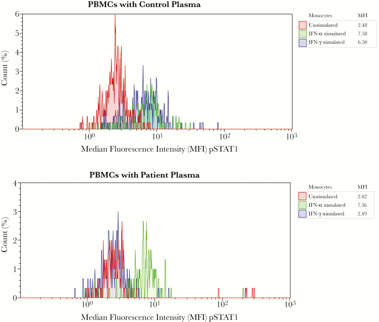
Figure 3.
Flow cytometry of a functional assay for detecting anti–interferon (IFN) γ autoantibodies. Healthy control peripheral blood mononuclear cells were surface stained with CD14-PC5 to identify monocytes and incubated with control (autologous) plasma and patient plasma in 3 conditions: unstimulated, IFN-α stimulated (as an internal control), and IFN-γ stimulated. Cells were stained for intracellular phosphorylated STAT1 (pSTAT1) with anti-STAT1 conjugated to Alexa Fluor 488 (Becton Dickinson), fixed, and then analyzed with flow cytometry. Median fluorescence intensities (MFIs) for pSTAT1 in CD14+ gated monocytes for each condition are compared between unstimulated and stimulated cells cultured in control and patient plasma. Red peak represents unstimulated cells; green peak, IFN-α–stimulated cells; and blue peak, IFN-γ–stimulated cells. Top plot (control plasma) shows increase in MFI, tabled under “MFI,” after stimulation with IFN-α and IFN-γ (blue and green peaks), and bottom plot (patient plasma) shows absence of pSTAT1 when stimulated with IFN-γ (blue peak), suggesting an inhibitor to IFN-γ in the patient’s plasma. This functional assay does not determine whether autoantibodies in the patient’s serum are targeting IFN-γ itself or IFN-γ receptors. Target-specific enzyme-linked immunosorbent assay was not performed for confirmation.

