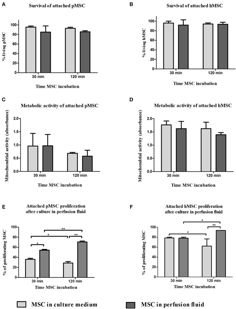
Figure 6.
Effect of perfusion fluid on attached MSC. Survival of attached pMSC (A) and hMSC (B) after 30 min in perfusion fluid. (C,D) Metabolic activity of attached pMSC (C) and hMSC (D) after 30 min in perfusion fluid measured by reduction of XTT. (E,F) Proliferation of attached pMSC (E) and hMSC (F) after 30 in perfusion fluid. Cells were trypsinized and re-seeded in a culture flask. Proliferation after 24 h was determined by CFSE fluorescence (n = 5). Results are shown as means ± SD. *p < 0.05; **p < 0.01.

