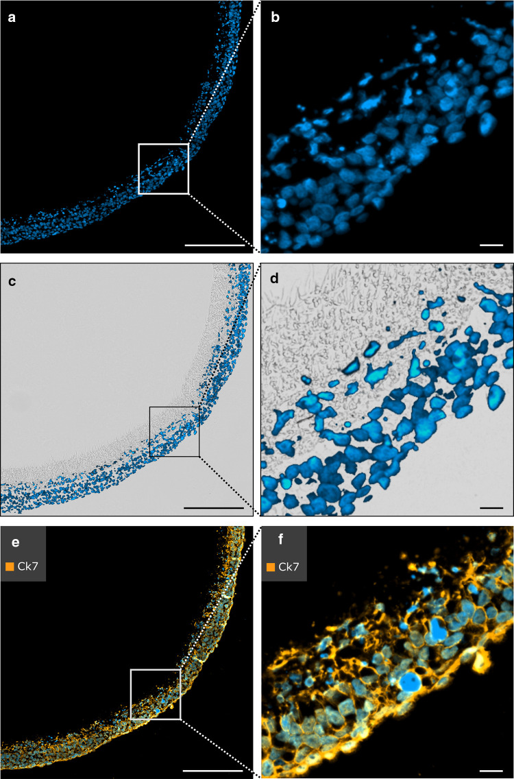
Fig. 5.
Immunofluorescent staining of cellularized PCL/PLA membranes cellularized with trophoblast cells. Sections were fixed in 3.7% PFA, embedded in low-melting-point paraffin (max. 50 °C), and antigen retrieval was performed with pepsin for 30 min. Nuclei were counterstained with DAPI (blue). a, b DAPI staining reveals successful cellularization of the PCL/PLA membrane. c, d Overlay image with fluorescence and bright field microscopy. Trophoblast cell migration into the PCL/PLA membrane is visualized with DAPI staining. e, f Sections are immunostained with anti-CK7. CK7 is used with Alexa Fluor 633 secondary antibody. Immunofluorescence demonstrates positive expression of CK7 in trophoblast cells. CK7 cytokeratin 7, PCL/PLA polycaprolactone/polylactide, PFA paraformaldehyde. Scale bars in a, c, e represent 200 µm, and those in b, d, f represent 20 µm

