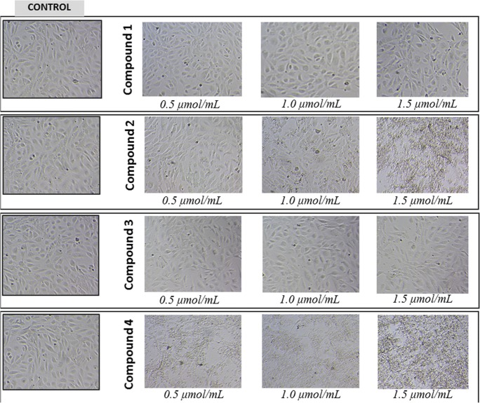
Fig. 8.
The effect of gadolinium complexes with iminodiacetic acid derivatives on morphology of HUVECs. HUVECs cultured in monolayer on 48-well plates were stimulated with indicated concentrations (0.5–1.5 μmol/mL) of compounds 1–4. Representative phase-contrast images are shown (magnification of 100 times). Compounds 1 and 3 over the entire concentration range did not contribute to the changes in cell morphology. Compound 2 at 1 μmol/mL caused cell membrane disintegration, while compound 4 even at the lowest concentration contributed to the lysis of cells

